
Sebutkan Struktur dan fungsi Trakea pada manusia Artikel Hiham.id
Mucoepidermoid carcinoma. Mucoepidermoid tumors are very rare tumors of the trachea and main bronchi and may be of low-grade or high-grade malignancy. On imaging, they present as a focal soft tissue endoluminal mass without distinguishing features from other airway malignancies ( Figure 7 ).
Trachea Anatomy & Function Trachea and Esophagus Location
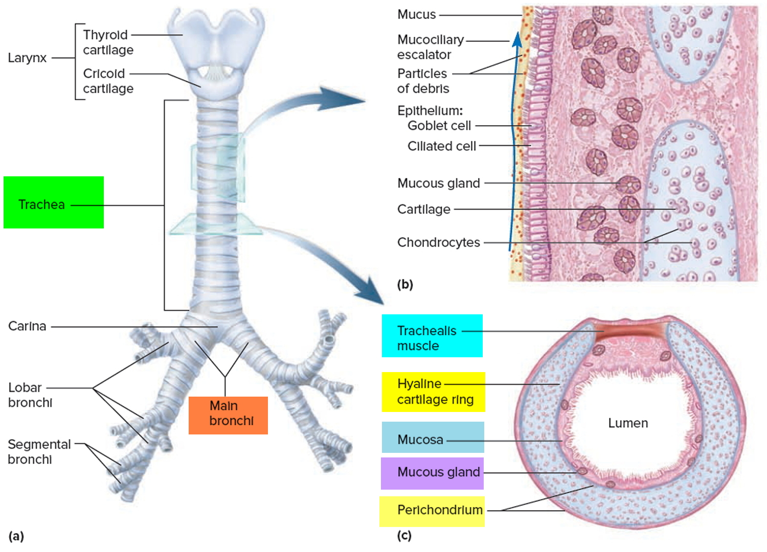
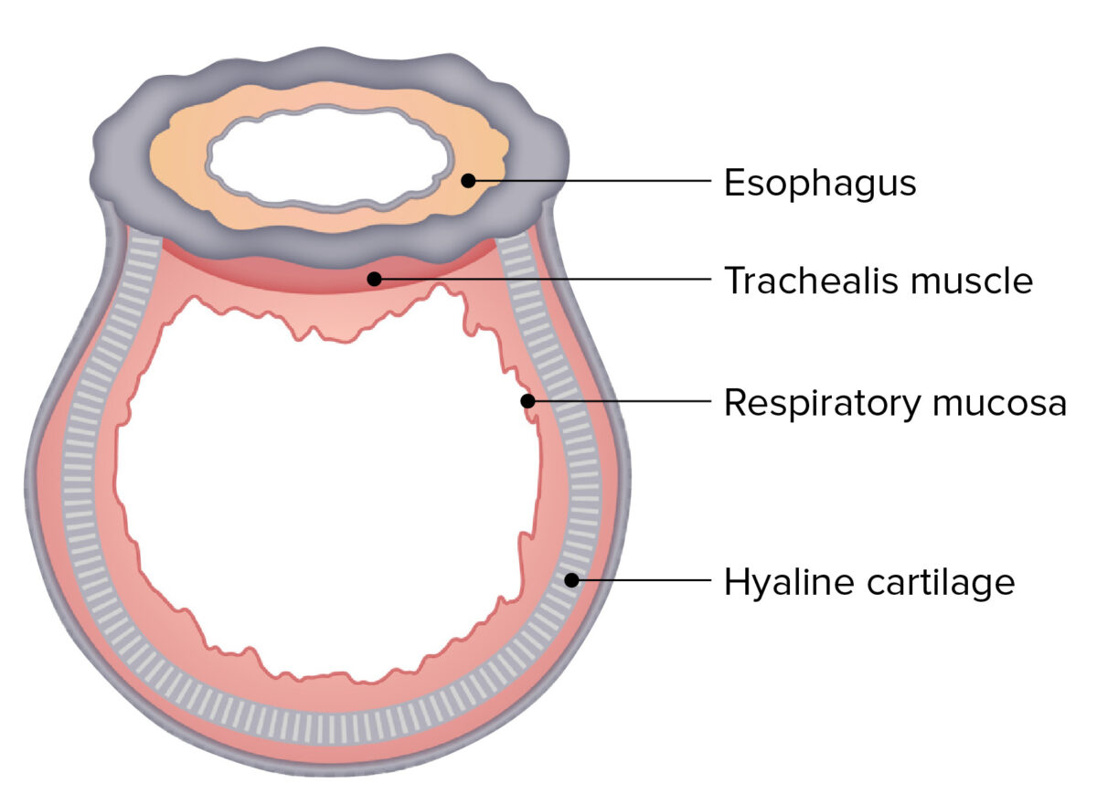
The trachea is a tube-shaped structure consisting of 15-20 D-shaped cartilage rings anterolaterally bridged by annular ligaments. The trachealis muscle (smooth muscle) encircles the trachea completely but is most prominent posteriorly due to the lack of cartilage 4. The trachea extends from the inferior margin of the cricoid cartilage (C6) and.

trachea in a body
This study aimed to investigate the effects of intravenous injection of iodine contrast agent on the tracheal diameter and lung volume. In this retrospective study, a total of 221 patients (71.1 ± 12.4 years, 174 males) who underwent vascular dynamic CT examination including chest were included. Unenhanced, arterial phase, and delayed-phase images were scanned. The tracheal luminal diameters.
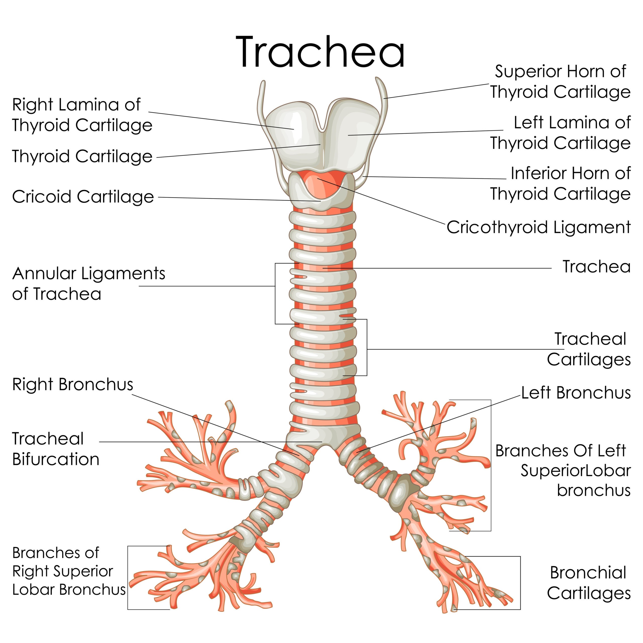
Trachea Diagram Labeled
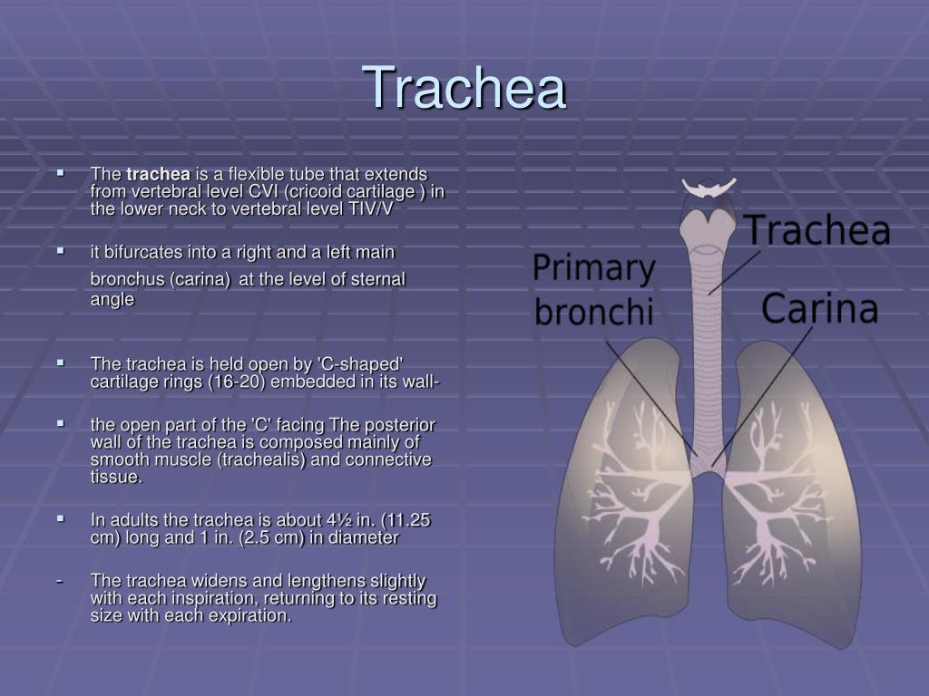
The trachea is a long tube that extends from the pharynx and larynx to the bronchi of the lungs. It typically has an inner diameter of about 25.4 millimeters (1.00 in) and a length of about 10 to 16 centimeters. The trachea commences at the lower border of the larynx, level with the sixth cervical vertebra, and bifurcates into the primary.

The Trachea or Windpipe Medika Life Understanding Human Anatomy
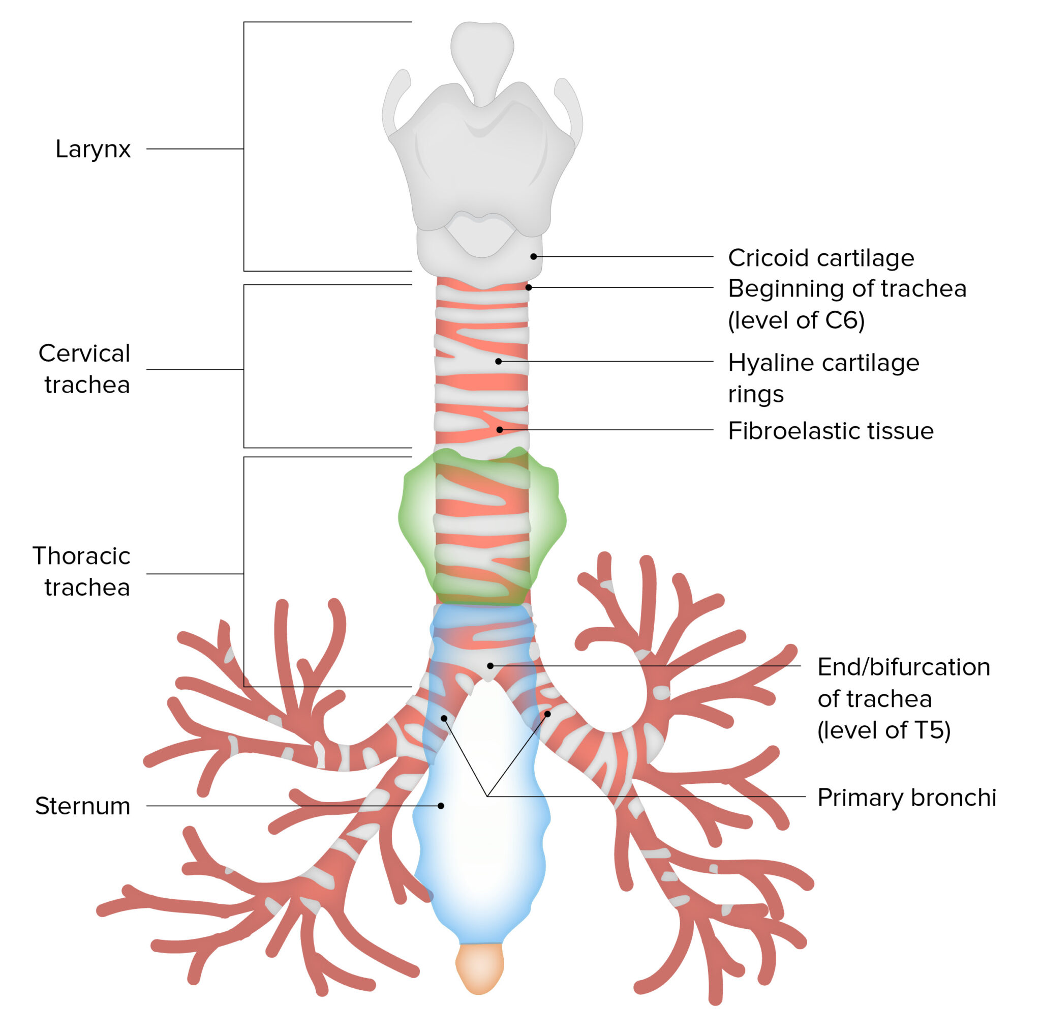
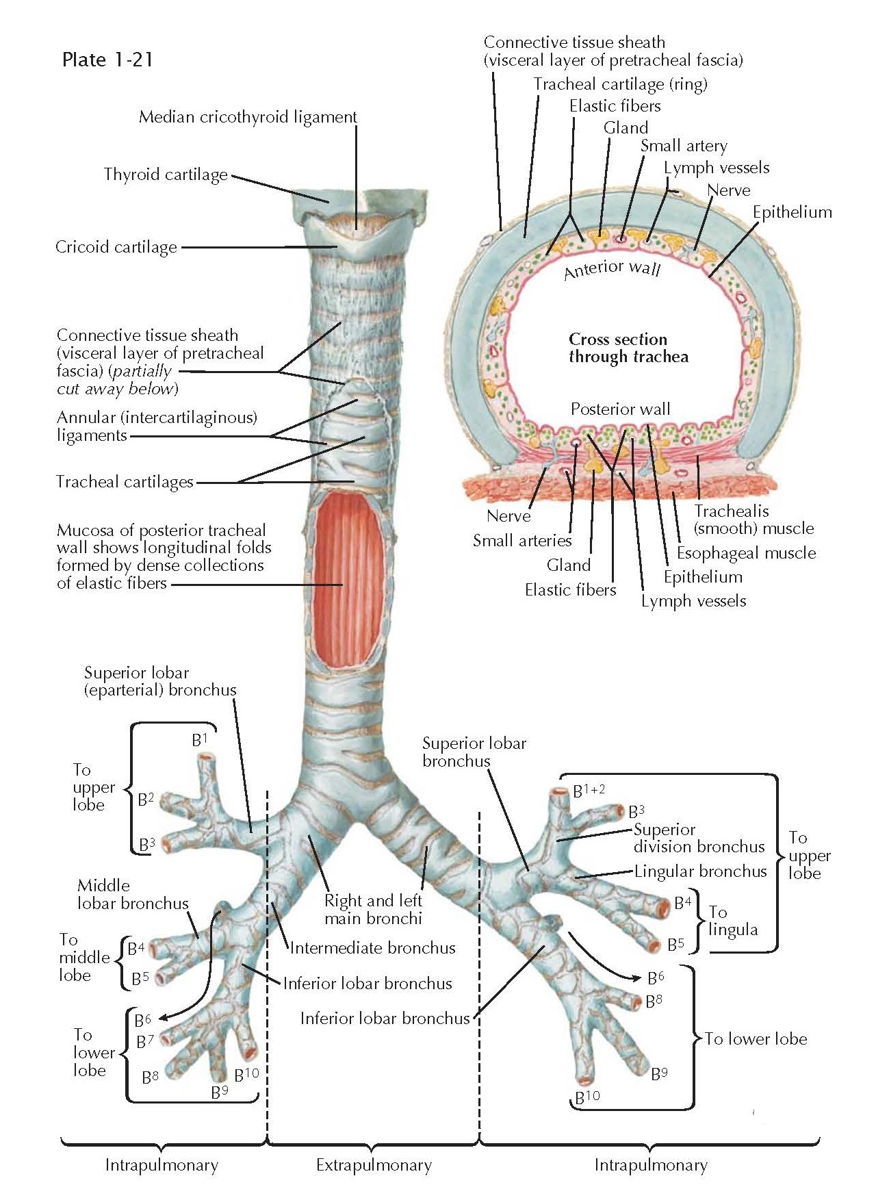
The trachea, or windpipe, is a 9-15 cm long fibrocartilaginous tube of the lower respiratory tract.It forms the trunk of the tracheobronchial tree, or pulmonary conducting zone.The trachea extends between the larynx and thorax, consisting of two parts; cervical and thoracic.It ends at the level of the sternal angle (T5) where it divides into two main bronchi, one for each lung.
Measurement of anteroposterior (1) and transverse (2) diameter of the
The upper limits of the coronal and sagittal diameters in men are 25 and 27 mm, respectively. In women, they are 21 and 23 mm, respectively. The lower limits for both dimensions are 13 and 10 mm for adult males and females, respectively. The trachea's diameter, from side-to-side, is from 2 to 2.5 cm, always greater in the male than in the female.
PPT Trachea and lungs PowerPoint Presentation, free download ID4779845
There are approximately two rings of cartilage per centimeter of trachea and each tracheal ring is an average of 4 mm in height. The wall of the trachea averages about 3 mm in thickness. The average external diameter of the trachea is 2.3 and 1.8 cm in the coronal and sagittal dimensions, respectively. Figure 2.

Anatomy of the Trachea, Carina, and Bronchi Thoracic Surgery Clinics
The trachea is a cartilaginous tube formed by a series of tracheal cartilages, joined together by annular ligaments (Figs. 4-13, 4-15 and 6-9).These cartilages give the trachea rigidity and prevent its collapse. In the mouse, the trachea has about 15 cartilages with an approximate internal diameter of 1.5 mm.The C-shaped cartilages are open dorsally.

Lower Respiratory System
Objective: To design and test a bench model of an intraluminal optical device capable of accurately measuring airway diameter. Design: A fresh porcine trachea divided longitudinally and affixed to a linear translation stage was used to simulate 20 tracheal diameters (18.3-30.3 mm). Tungsten-halogen light was dispersed across the luminal surface by a diffraction grating.

Benign and Malignant Disorders of the Trachea Current Diagnosis
Trakea memiliki panjang sekitar 10cm pada wanita dan panjang cm pada pria. Memiliki diameter anterior-posterios rata-rata mm, diameter transversal 18mm. Trakea memanjang mulai dari bawah laring, setinggi vertebra sevikalis 6 hingga vertebra torakalis 4. Trakea terbagi menjadi dua bronkus, yaitu bronkus utama kanan dan kiri.
Trachea Anatomy Concise Medical Knowledge
The function of the trachea is to be the main passageway for air to pass from the upper respiratory tract to the lungs. As air flows into the trachea during inhalation, it is warmed and moisturized before entering the lungs. Most particles that enter the airway are trapped in the thin layer of mucus on the trachea walls.
Trachea Anatomy Concise Medical Knowledge
Tracheal diameter varies widely in normal subjects. In normal men, tracheal diameter averages 19.5 mm, with a range of 13 to 25 mm (mean ± 3 SD) in the coronal plane and 13 to 27 mm in the sagittal plane. In women, tracheal diameter is slightly smaller, averaging 17.5 mm and ranging from 10 to 21 mm in the coronal plane and 10 to 23 mm in the.

Relationship between the trachea and esophagus highlighting the
Saber-sheath trachea refers to a diffuse coronal narrowing of the intrathoracic portion of the trachea with the concomitant widening of the sagittal diameter. It is not uncommon and is pathognomonic for chronic obstructive pulmonary disease (COPD) 1 . The sagittal:coronal diameter is over 2:1 2 and the extra-thoracic portion of the trachea is.

Axial image with measurement of the width (diameter) of trachea
Structure. An adult's trachea has an inner diameter of about 1.5 to 2 centimetres (1 ⁄ 2 to 3 ⁄ 4 in) and a length of about 10 to 11 cm (4 to 4 + 1 ⁄ 4 in), wider in males than females. The trachea begins at the lower edge of the cricoid cartilage of the larynx at the level of sixth cervical vertebra (C6) and ends at the carina, the point where the trachea branches into left and right.
Cancer De Traquea
Assuming a normative range that encompasses three standard deviations from the mean or 99.7% of the normal population, the upper limits of normal for coronal and sagittal diameters, respectively, in men aged 20-79, are 25 mm and 27 mm; in women, they are 21 mm and 23 mm, respectively. The lower limit of normal for both dimensions is 13 mm in.
Trachea Diagram Labeled
The trachea or windpipe is a cartilaginous and membranous tube, extending from the lower part of the larynx, on a level with the sixth cervical vertebra, to the upper border of the fifth thoracic vertebra, where it divides into the two bronchi, one for each lung. The trachea is nearly but not quite cylindrical, being flattened posteriorly; it measures about 11 cm. in length; its diameter, from.