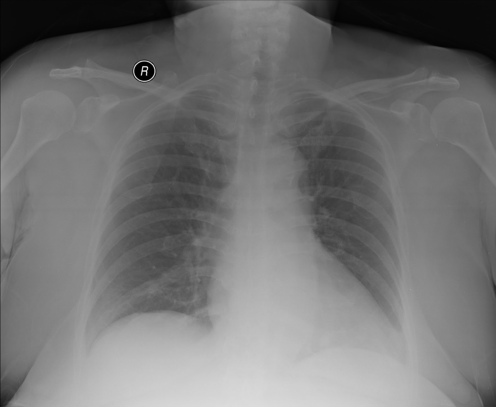
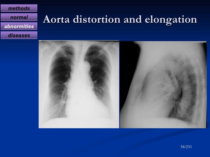
Chest X Ray Elongatio aorta Radiology Imaging
Hasil yang akan diamati adalah besar jantung dan kejadian elongasi aorta. Hasil pengamatan akan dianalisa korelasinya dengan tekanan darah menggunakan uji korelasi Spearman. Hasil : Terdapat hubungan bermakna antara tekanan darah dengan besar jantung (p=0,007), dan memiliki korelasi positif yang rendah (nilai korelasi 0,298) .

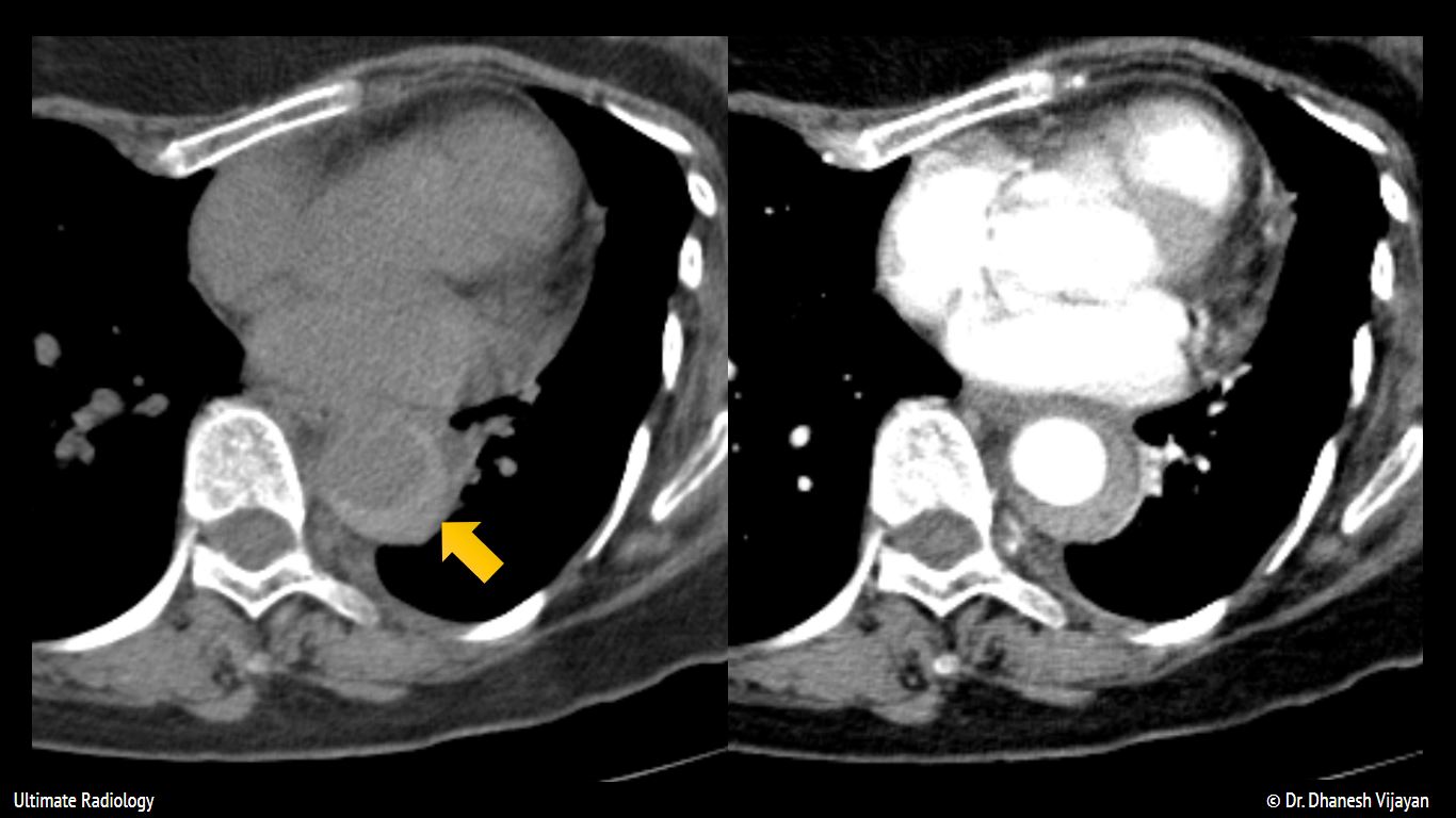
Aortic Dissection CT Scan
The normal aortic ageing process is accompanied by gradual luminal dilatation and reduction of vessel compliance. However, the influence of age on longitudinal aortic dimensions and geometry has not been well studied. This study aims to describe the normal evolution of aortic length and shape throughout life. Methods: A total of 210 consecutive.

Elongasi Aorta PDF
Penelitian ini bertujuan untuk mengidentifikasi hubungan kejadian elongasi aorta melalui gambaran foto toraks dengan hipertensi. Metode : Studi penelitian menggunakan pendekatan cross sectional pada 104 pasien untuk mengidentifikasi usia, jenis kelamin, dan nilai tekanan darah dari rekam medis pasien.. Hanna Marsinta Uli Bagian Radiologi.

Aortic coarctation Image
Aortic aneurysms are focal or diffuse dilatations of the aorta involving all three aortic wall layers. Most aneurysms are caused by atherosclerosis, whilst trauma, infection, and genetic syndromes are other causes. The broad term aortic aneurysm is usually reserved for pathology discussion. More specific anatomic and radiologic discussion is based on the location of the aneurysm:

Echocardiography Of The Ascending Aorta Steve Gallik
2Bagian Radiologi Fakultas Kedokteran Universitas Sriwijaya/. elongasi aorta sebanyak 9,792 kali pada pasien hipertensi dibandingkan pasien yang tidak hipertensi (Tabel 3).
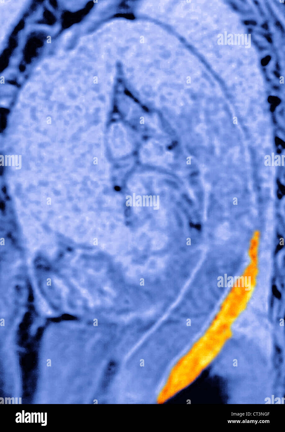
AORTIC DISSECTION, MRI Stock Photo Alamy
Differential diagnosis. senile ectasia. hypertension. post-stenotic dilatation, e.g. bicuspid aortic valve. thoracic aortic aneurysm. atherosclerosis (usually descending thoracic aorta) collagen disorders. Marfan syndrome. Ehlers-Danlos syndrome (classically involves sinuses of valsalva)
Chest Xray Interpretation A Structured Approach Radiology OSCE
Prosedur radiologi memungkinkan dokter melihat bagian dalam tubuh dengan sangat detail, sehingga dapat membantu dalam menegakkan diagnosis elongasi aorta dengan akurat. Hasil klinis yang lebih akurat dapat memfasilitasi penanganan dan pengobatan yang tepat, termasuk strategi operasi dan pemberian obat untuk menghindari pecahnya aorta.

Aortic dissection echocardiography and ultrasound wikidoc
Latar belakang : Hipertensi adalah suatu peningkatan tekanan darah di luar batas normal. Kondisi ini dapat menyebabkan berbagai komplikasi kesehatan termasuk perubahan pada struktur pembuluh darah. Perubahan struktural pada aorta berupa pemanjangan aorta disebut elongasi aorta yang terlihat melalui pemeriksaan foto toraks. Penelitian ini bertujuan untuk mengidentifikasi hubungan kejadian.
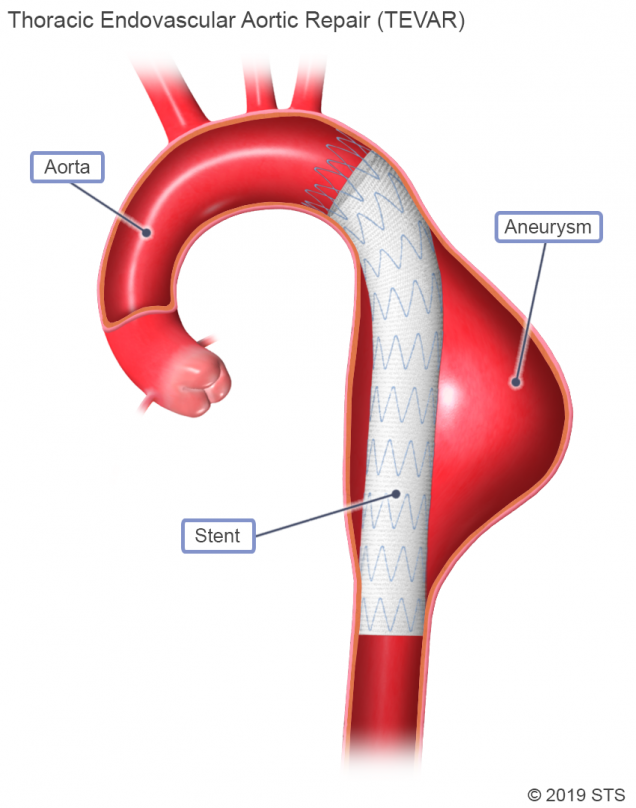
Thoracic Aortic Aneurysm The Patient Guide to Heart, Lung, and Esophageal Surgery
The aorta, the great artery, is the largest artery of the human body and carries oxygenated blood ejected from the left ventricle to the systemic circulation. It is divided into: thoracic aorta. ascending aorta; aortic arch; descending aorta; abdominal aorta; It has branches from each section and gradually tapers down to its termination where it bifurcates into the common iliac arteries.

How to Measure the Aorta Using MRI A Practical Guide Hout 2020 Journal of
1. Introduction. Aortic morphology has been reported to be associated with age. A study of 123 subjects without thoracic aorta pathology or surgery found that the aortic diameter enlarges, the posterior arch elongates, the tortuosity index decreases, and the attachment zone angle is larger in older people [].Age had a correlation coefficient of 0.61 with arch length (p < 0.01) in the study.
Aortic Stenosis a Tight Aortic Valve A Comprehensive Patient Guide • MyHeart
Objectives Differentiation between normal and abnormal features of vascular ageing is crucial, as the latter is associated with adverse outcomes. The normal aortic ageing process is accompanied by gradual luminal dilatation and reduction of vessel compliance. However, the influence of age on longitudinal aortic dimensions and geometry has not been well studied. This study aims to describe the.

Aortic Dissection Imaging Images
Elongasi aorta dapat terlihat melalui teknik radiologi seperti sinar-X, MRI, CT scan, atau ultrasonografi. Elongasi aorta dapat menimbulkan gejala pada beberapa orang seperti nyeri dada, kesulitan bernapas, dan sesak napas. Komplikasi yang dapat timbul akibat kondisi ini adalah pecahnya aorta, stroke, atau serangan jantung.
Aortic Dissection Imaging Images
gambaran radiologi elongasi aorta by rizka9rinintia9sari

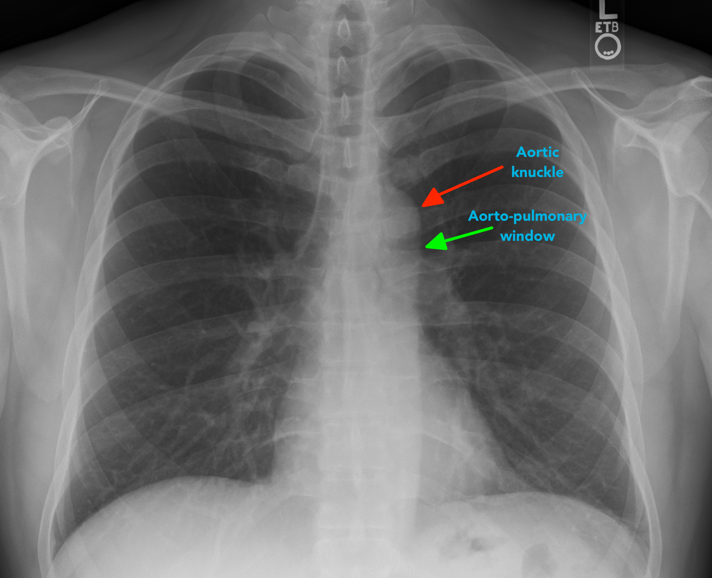
(PDF) Relation between aortic knob width and subclinical left ventricular dysfunction in
Hasil studi didapatkan hubungan bermakna antara usia dengan elongasi aorta (p-value = 0.001) dengan resiko peningkatan panjang aorta 1,065 kali per tahun. Berdasarkan hasil studi ini dapat.
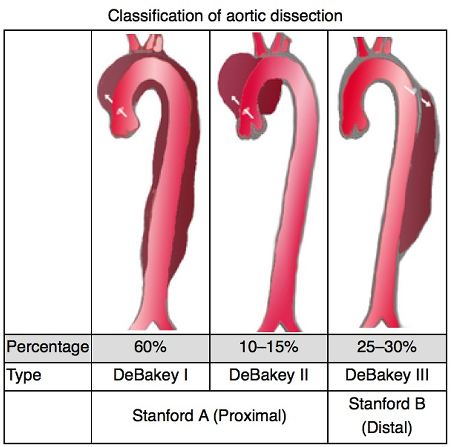
Aortic Dissection Type A And B Symptoms, Causes, Treatment
The elongated aorta It is the imaging finding in which the aorta, the main artery of the human body, is observed longer than normal. It was initially described only in thoracic radiology, but the term was extrapolated to other studies that include images, such as CT scans, MRIs or catheterizations. In chest radiographs taken anteroposterior or.
Diagnostic radiology of cardiovascular 2009
Hasil MCU saya dari radiologi thorax kesannya Aorta Elongasi dan Curiga Dilatasi. Info lainnya al. usia: 57 th, lingkar perut: 99, tensi: 171/90, kolesterol: 255, asam urat: 8,6, gampang lelah/lemas, kurang keseimbangan serta kadang-kadang sesak nafas. Mohon advis dokter untuk tindak lanjut dan pengobatan yang diperlukan.
