
Xray of shoulder joint A/P & Lateral View Proper position of shoulder joint.👀 YouTube
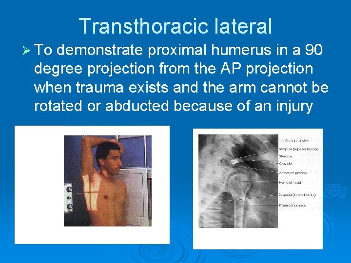
The inferosuperior axial view also known as a Lawrence view of the shoulder is a modified axial projection best utilized with supine patients. It is an orthogonal projection to the AP view and replaces the lateral shoulder projection. Indications
Gudang Medis Teknik Radiografi Shoulder Joint
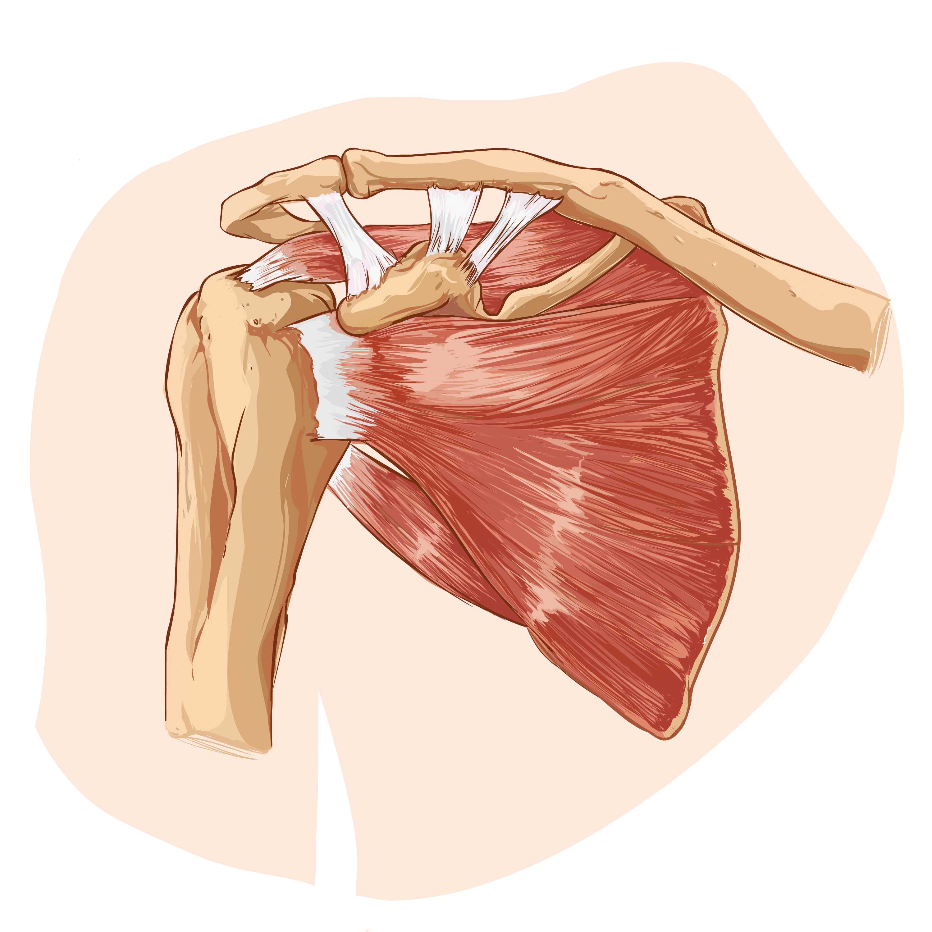
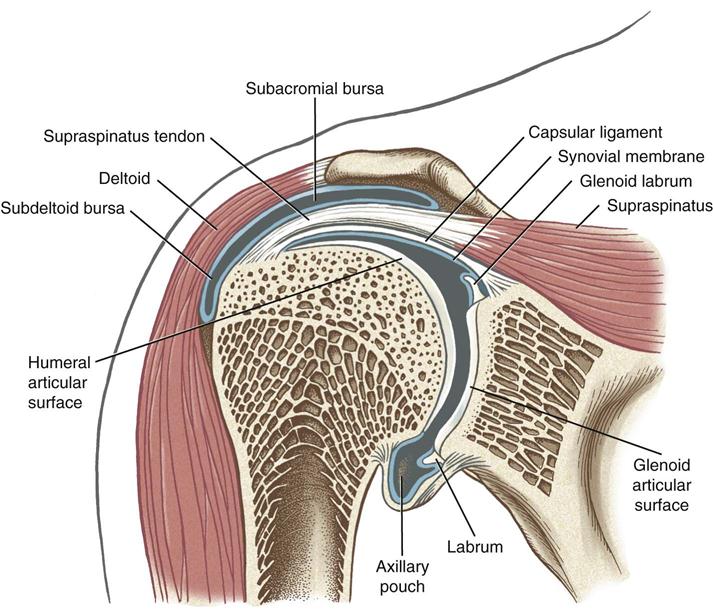
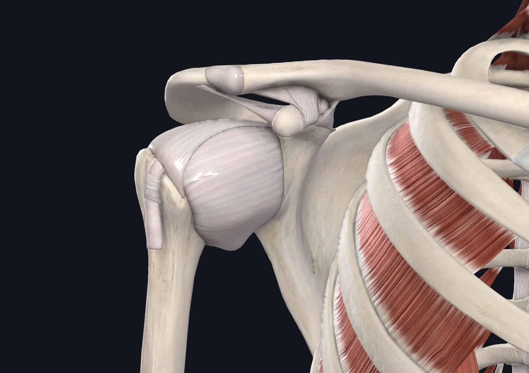
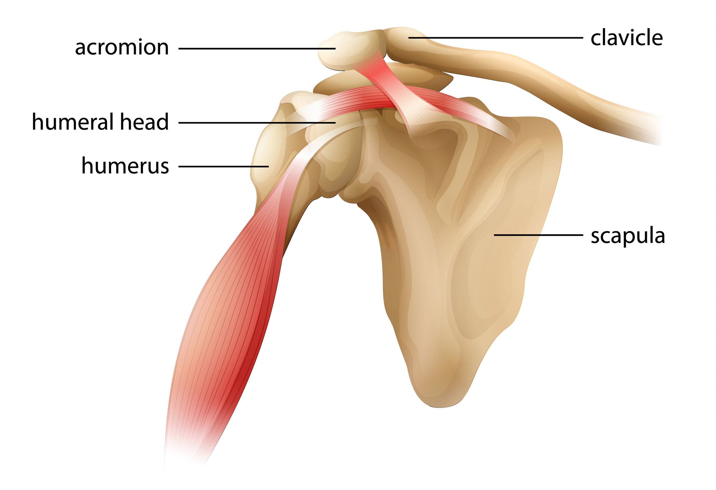
The glenohumeral, or shoulder, joint is a synovial joint that attaches the upper limb to the axial skeleton. It is a ball-and-socket joint, formed between the glenoid fossa of scapula (gleno-) and the head of humerus (-humeral). Acting in conjunction with the pectoral girdle, the shoulder joint allows for a wide range of motion at the upper.

Figure 6 from Radiographic evaluation of the shoulder. Semantic Scholar
Prosedur pemeriksaan radiografi shoulder joint pada klinis trauma dilakukan dengan teknik yang bervariasi dalam beberapa jurnal. Penelitian ini bertujuan untuk menjelaskan pemilihan proyeksi pemeriksaan radiografi shoulder joint yang optimal pada kasus trauma bahu berdasarkan literature review Jenis penelitian kualitatif deskriptif dengan.
the shoulder joint On Target Publications
The results of test functions are analyzed based check list of respondents about the value of work tools.Results: The results of design tools fixation on radiographic examination shoulder joint.
Shoulder Glenohumeral Joint AP shoulder girdle Three projections
the level of the glenohumeral joint on the posterior aspect of the patient (5 cm below the top of the shoulder) central to the medial scapula border. collimation. laterally to include the skin margin. medially to cover the entirety of the medial scapula. superior to the skin margin. inferior to the inferior angle of the scapula.

Conventional radiographs of the shoulder. (A) Anteroposterior (AP) view... Download Scientific
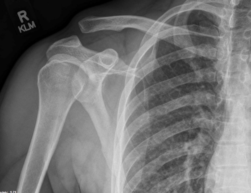
The shoulder AP view is a standard projection that makes up the two view shoulder series. The projection demonstrates the shoulder in its natural anatomical position allowing for adequate radiographic examination of the entire clavicle and scapula, as well as the glenohumeral, acromioclavicular and sternoclavicular joints of the shoulder girdle.
Acromioclavicular Joint Separation Undergraduate Diagnostic Imaging Fundamentals
Teknik Pemeriksaan Radiografi Shoulder Dipos oleh Rini pada Juni 13, 2021 Proyeksi : AP (External Rotation) Kaset : ukuran 24 x 30 cm kV : 70 ± 5 mAs : 6 FFD : 100 cm Posisi Pasien : Posisi pasien erect atau supine. (Posisi erect biasanya mengurangi sakit pada pasien) Posisi Obyek : Putar tubuh ke shoulder yang sakit sehingga menempel ke kaset.

Shoulder Joint Anatomy Pictures and Information
Proyeksi Pemeriksaan Shoulder Joint ada 9 yaitu AP Eksorotasi AP Endorotasi AP Neutral AP External Rotasi :Shoulder (Non Trauma) AP-Internal Rotasi : Shoulder (Non Trauma) Inferosuperior Axial : Shoulder (Non Trauma) Posterior Obliq - Glenoid cavity : Shoulder (Non Trauma) Tangensial-Intertubercular (Bicipital) Groove : Shoulder (Non Trauma)
Shoulder Musculoskeletal Key
SARAN Berdasarkan hal di atas penulis Dalam pemeriksaan radiografi berpendapat bahwa setiap proyeksi shoulder joint pada kasus trauma bahu memiliki kelebihan dan kekurangannya dapat menggunakan Scapular Y View masing-masing dan oleh karena itu sebagai proyeksi tambahan untuk penulis memiliki anggapan bahwa pada mengevaluasi dislokasi, apabila.
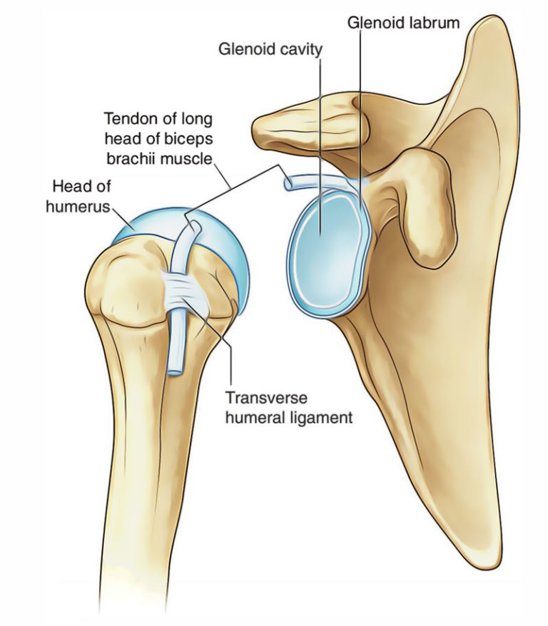
Shoulder Joint (Glenohumeral Joint) Earth's Lab
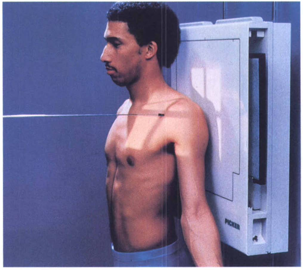
Latar Belakang : Menurut Lampignano dan Kendrick (2018) proyeksi, yang digunakan pada pemeriksaan radiografi shoulder joint pada pasien trauma yaitu Anterior posterior (Neutral Rotation), Trans thoracic Lateral, PA Oblique Scapular Y lateral), Proyeksi Tangensial (Supraspinatus Outlet), AP Apical Oblique.

Normal shoulder Image
PDF | On Jul 15, 2019, Agustina Dwi Prastanti and others published PENGARUH VARIASI ROTASI TUBUH TERHADAP INFORMASI ANATOMI PADA PEMERIKSAAN RADIOGRAFI SHOULDER JOINT METODE SCAPULAR Y VIEW | Find.

Gudang Medis Teknik Radiografi Shoulder Joint
Dislokasi sendi merupakan keadaan di mana tulang- tulang yang membentuk sendi tidak lagi berhubungan secara anatomis. proyeksi yang digunakan dalam kasus dislokasi ini yaitu AP dan Lateral.Tujuan penelitian ini yaitu mengetahui teknik radiografi pada pemeriksaan shoulder joint dengan kasus dislokasi dan mengetahui informasi anatomi proyeksi ante.
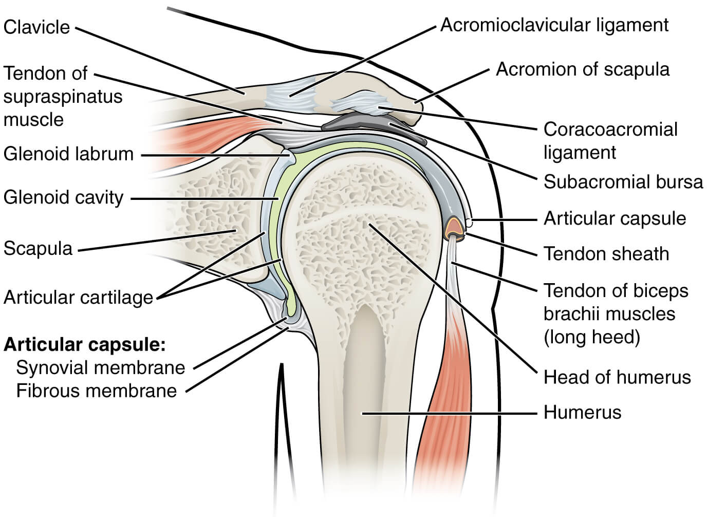
Glenohumeral Joint WikiMSK
Dislokasi sendi merupakan keadaan di mana tulang- tulang yang membentuk sendi tidak lagi berhubungan secara anatomis. proyeksi yang digunakan dalam kasus dislokasi ini yaitu AP dan Lateral.Tujuan penelitian ini yaitu mengetahui teknik radiografi pada pemeriksaan shoulder joint dengan kasus dislokasi dan mengetahui informasi anatomi proyeksi ante.
The Complete Guide to Shoulder Instability Labs
Hasil penelitian menunjukkan proyeksi pemeriksaan yang optimal pada pemeriksaan shoulder joint pada kasus trauma bahu adalah Anteroposterior (AP) dikarenakan menampakkan anatomi shoulder joint secara keseluruhan sehingga dapat mendiagnosa apabila terdapat dislokasi atau fraktur kemudian Scapular Y View sebagai proyeksi tambahan untuk mengevaluas.

Glenohumeral Joint Imaging Musculoskeletal Key
Kesimpulan : Penulis menyimpulkan bahwa proyeksi yang digunakan untuk pemeriksaan radiografi shoulder joint pada kasus dislokasi yaitu proyeksi modifikasi rotasi tubuh 50° scapular Y view, anteroposterior (AP) dan modifikasi proyeksi axillary view, Anteroposterior (AP) dan scapular Y view. Actions (login required) View Item
Shoulder Joint Pain Causes & Treatment Dr. Chris Homan
Abstract Latar belakang : Menurut Lampignano dan Kendrick (2018) pemeriksaan radiografi shoulder joint pada pasien dengan klinis dislokasi menggunakan proyeksi AP Neutral Rotation, PA Oblique, dan Transthoratic Lateral. Pemeriksaan radiografi shoulder joint di Instalasi Radiologi RST.
