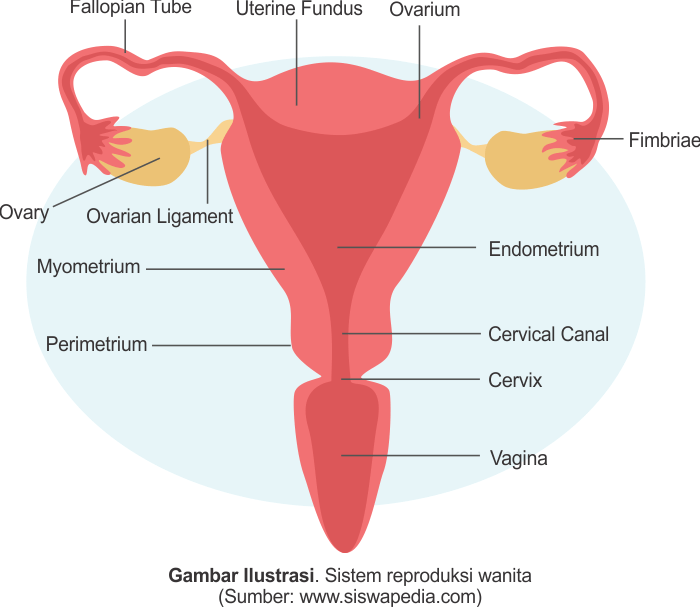
Fungsi Uterus atau Rahim Pada Sistem Reproduksi Wanita Siswapedia
Immediately postpartum, the uterine fundus is palpable at or near the level of the maternal umbilicus. Thereafter, most of the reduction in size and weight occurs in the first 2 weeks, at which time the uterus has shrunk enough to return to the true pelvis. Over the next several weeks, the uterus slowly returns to its nonpregnant state.

PPT Feminina PowerPoint Presentation, free download ID3305428
Pengertian dari fundus uteri adalah bagian dari uterus proksimal yang berada diatas muara tuba uterina yang mirip dengan kubah. Pada bagian ini tuba falloppii masuk ke uterus. Bagian dari fundus uteri ini juga dapat menjadi tempat perlengkatan plasenta di dalam rahim. Sedangkan pengertian korpus uteri adalah bagian uterus yang utama dan terbesar.

Coronal view of the fundus of the uterus on transvaginal ultrasound... Download Scientific Diagram
The uterus is a muscular, hollow organ in the female pelvis that is approximately 5 cm wide, 8 cm long, and 4 cm thick with a volume of 80 to 200 mL. A physiologically normal uterus typically lies in a position of anteversion (tilts forward at the cervix) and anteflexion (tilts forward at the isthmus). The uterus is situated posterior to the bladder, anterior to the rectum, and consists of.

Anatomy Of Uterus Anterior View
Introduction. Postpartum hemorrhage (PPH) is one of the leading causes of maternal morbidity and mortality worldwide 1, 2.The initial treatments for PPH are to administer uterotonic agents, uterine fundal massage, or bi-manual uterine compression 1, 2.Intrauterine gauze tamponade or an intrauterine balloon catheter may be employed in some instances 1, 2.

Gambar Tinggi Fundus Uteri
Temukan fundus Anda. Kemudian, temukan fundus (bagian 'atas' uterus) Anda dengan merasakan di dekat pusar. Buat otot perut rileks saat Anda memijit lembut area di atas dan di bawah pusar. Rasakan 'bukit' yang samar-samar di bawah kulit - ini adalah fundus Anda.

Uterine fibroids The Lancet
The uterus, also known as the womb, is an about 8 cm long hollow muscular organ in the female pelvis and lies dorsocranially on the bladder.It consists of several anatomical parts, such as the cervix, isthmus, and body. While its anatomy sounds simple, its histology is more complicated. It consists of three major layers, but the exact histological structure depends upon the state - if it is in.
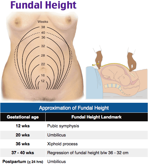
Image Fundal height, fundus, uterus, pregnancy
Kesimpulan. Tinggi fundus uteri atau TFU membantu memberikan informasi terkait perkembangan, ukuran, posisi, hingga masalah pada janin. Pengukuran ini mulai bisa dilakukan setelah usia kehamilan 20 minggu, tetapi tidak lagi efektif saat usia kehamilan 36 minggu. TFU yang normal adalah sesuai usia kehamilan Anda dengan selisih kurang-lebih 2 cm.
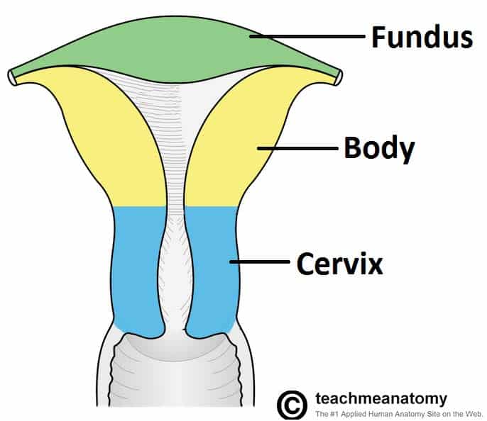
The Uterus Structure Location Vasculature TeachMeAnatomy
The fundus of uterus, also called the uterine fundus, refers to the dome-shaped, rounded superior part of the body of the uterus that lies above the opening of the uterine tubes.. It consists of a thick layer of smooth muscle tissue, the myometrium and forms the superior border of the uterine cavity. The uterine fundus is typically inclined slightly forward, creating an angle between the body.
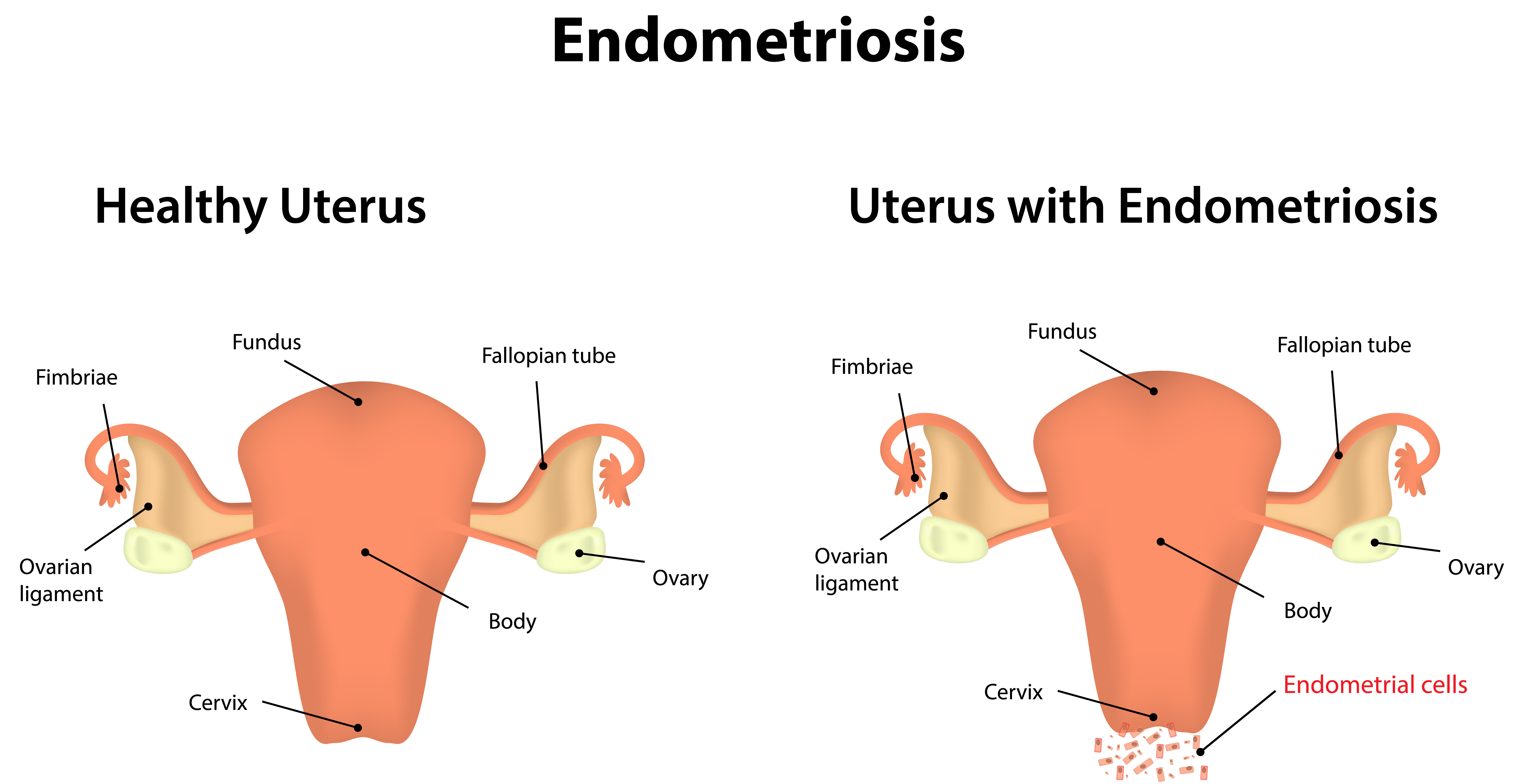
What Is Endometriosis?
Rahim atau uterus adalah salah satu organ kompleks dari sistem reproduksi wanita. Bentuk dari uterus seperti buah pir terbalik yang terletak di antara kandung kemih dan rektum. Sementara ukuran panjang uterus 7-7,5 cm dan lebar 5 cm dengan ketebalan sekitar 2,5 cm. Beratnya mencapai 60 gr dengan tebal dinding sekitar 1,25 cm.
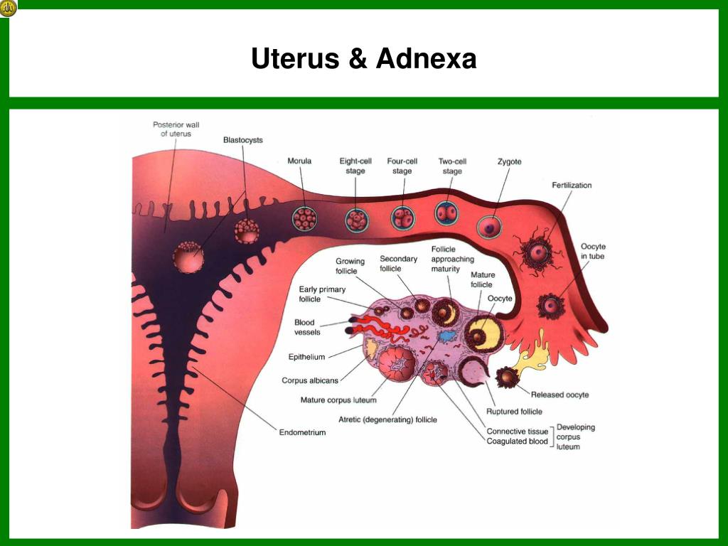
Uterus Adnexa Anatomy
A uterine inversion is a rare event, complicating about 1 in 2000 to 1 in 23,000 deliveries. Ironically, most are seen with "low-risk" deliveries. The incidence is 3-times higher in India as compared to the United States. The incidence of uterine inversion has decreased 4-fold after the introduction of active management during the third stage.
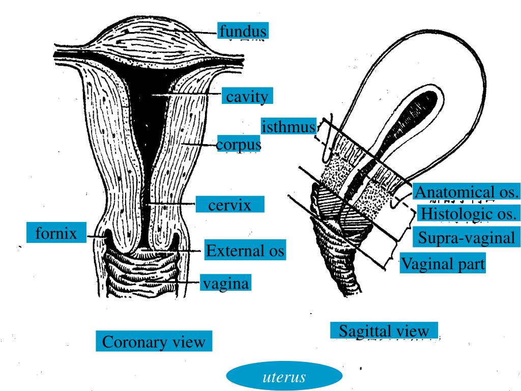
PPT Reference PowerPoint Presentation, free download ID1396839
Fundus berarti titik tertinggi, sedangkan uteri berarti rahim (uterus). Jadi, fundus uteri adalah titik tertinggi dari rahim. Tinggi fundus uteri merupakan jarak antara titik simfisis pubis dan fundus uteri yang biasanya dilakukan oleh dokter atau bidan.
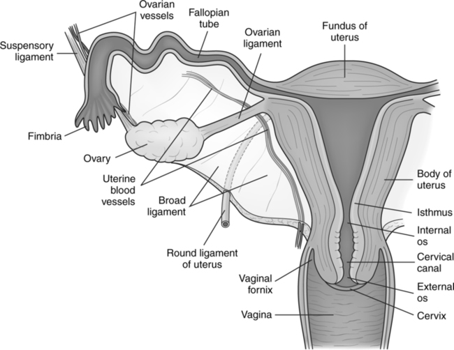
Uterus Side View Diagram
Uterine atony refers to the corpus uteri myometrial cells inadequate contraction in response to endogenous oxytocin that is released in the course of delivery. It leads to postpartum hemorrhage as delivery of the placenta leaves disrupted spiral arteries which are uniquely void of musculature and dependent on contractions to mechanically squeeze them into a hemostatic state. Uterine atony is a.
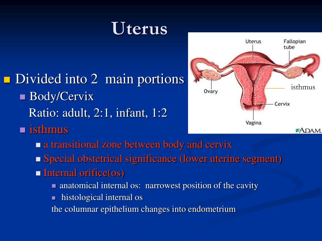
PPT Reference PowerPoint Presentation, free download ID1396839
The uterus is a hollow, pear-shaped organ that is responsible for a variety of functions, such as gestation (pregnancy), menstruation, and labor and delivery. On a coronal cut section, its cavity has an inverted triangle shape. Sometimes the development in utero may be incomplete; this is called a Mullerian anomaly and can lead to many variants, ranging from a uterine septum to uterine.
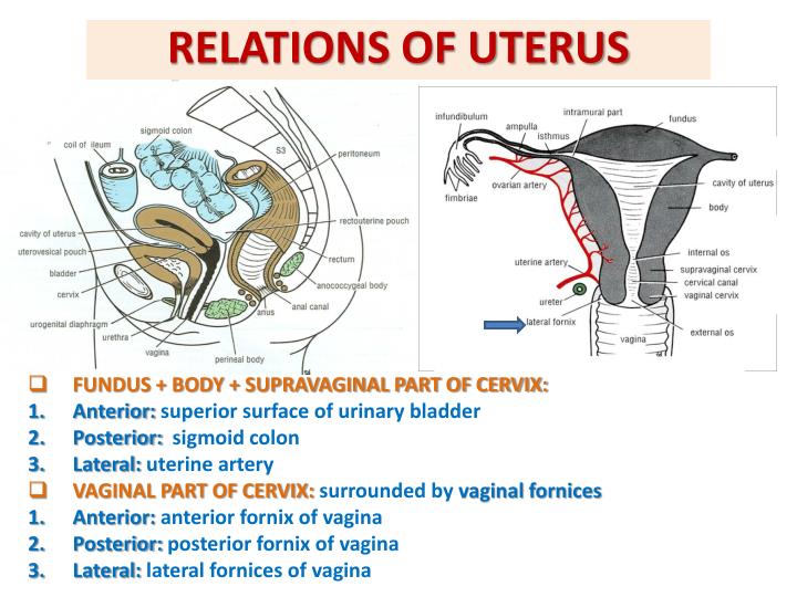
Posterior Uterus Anatomy
Uterus adalah organ dalam sistem reproduksi wanita yang terletak di dalam pelvis. Fungsi dari uterus adalah sebagai tempat penampungan sel telur yang telah dibuahi hingga membentuk janin yang siap untuk dilahirkan.. Fundus merupakan bagian paling atas dan terluas dari uterus yang terhubung ke saluran tuba. Pada bagian ini, tuba falopi akan.

Uterus Anatomy,Function, Inverted, Tipped & Transplantation
Penjelasan tentang Fungsi Uterus pada Wanita. Reproduksi. Ditinjau oleh dr. Verury Verona Handayani 11 Januari 2021. Halodoc, Jakarta - Uterus (juga disebut rahim) adalah organ otot berbentuk buah pir terbalik dari sistem reproduksi wanita yang terletak di antara kandung kemih dan rektum.

FEMALE INTERNAL ORGAN Pharmahelp
If the length of the entire uterus (including the cervix) is required (e.g. at preoperative evaluation), the sum of the total length of the uterine corpus (d1) and the cervical length should be reported. d1 is calculated as the sum of the fundal length (from the fundal serosal surface of the uterus to the fundal tip of the endometrial cavity.